
Mediastinal Lymph Node Quantification (LNQ): Segmentation of Heterogeneous CT Data
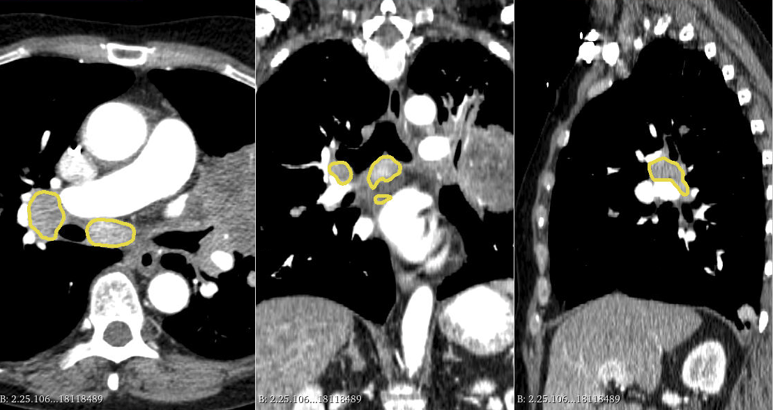
Example of expert annotations of mediastinal lymph nodes on a
contrast-enhanced CT scan of the type to be used in the challenge.
🎬 Introduction
Accurate lymph node size estimation is critical for staging cancer patients, initial therapeutic management, and in longitudinal scans, assessing response to therapy. Current standard practice for quantifying lymph node size is based on a variety of criteria that use unidirectional or bidirectional measurements on just one or a few nodes, typically on just one axial slice. But humans have hundreds of lymph nodes, any number of which may be enlarged to various degrees due to disease or immune response. While a normal lymph node may be approximately 5 mm in diameter, a diseased lymph node may be several cm in diameter. The mediastinum, the anatomical area between the lungs and around the heart, may contain ten or more lymph nodes, often with three or more enlarged greater than 1 cm. Accurate segmentation in 3D would provide more information to evaluate lymph node disease. Full 3D segmentation of all abnormal lymph nodes in a scan promises to provide better sensitivity to detect volumetric changes indicating response to therapy.
Machine learning models for medical image segmentation have shown remarkable progress in recent years, but we know of no tools available to fully quantify lymph nodes, probably in part because of a lack of pixel-level ground truth annotations for training. In our view, there are two critical roadblocks that hamper the development of automated lymph node identification and segmentation tools. First, lymph nodes are often within the intensity profile of normal soft tissue and have ill-defined borders, which makes them difficult to identify without medical training. This problem is further complicated by the fact that nodes can become confluent, leading to larger matted structures which have no characteristic shape. Their presentation across subjects can vary significantly, making it difficult to scale from small datasets to robust tools. Second, since there is frequently more than one diseased node per case, and manual annotation is time-consuming, there are no pre-existing clinical use cases where cases are being fully annotated. These critical issues make lymph node segmentation an ideal candidate for weakly-supervised segmentation techniques. Weakly-supervised learning has recently garnered strong interest in the medical imaging community, and the development of robust weakly-supervised techniques will enable machine-learning practitioners to leverage the weak annotations that are captured every day during real-world clinical operations.
While a large variety of weakly-supervised techniques have been proposed for image segmentation, most of these techniques have been validated either on private datasets or by artificially creating partial annotations from fully annotated datasets. The proposed LNQ2023 challenge will provide the first medical benchmark for evaluating the performance of weakly-supervised segmentation techniques for lymph node segmentation on a large dataset part of a clinical trial. The LNQ2023 Challenge invites members of the MICCAI community to develop a system that automatically segments mediastinal lymph nodes in CT scans. The LNQ2023 Challenge training dataset will consist of a unique set of high-quality pixel-level annotations of one or more clinically relevant lymph nodes in a dataset part of cancer clinical trials. The goal will be to segment all lymph nodes larger than 1 cm in diameter in the mediastinum. Participants will be provided with a subset of cases that are partially annotated (i.e. one node out of five), and evaluation of the algorithms will be performed on a distinct dataset that is fully annotated (i.e. all clinically relevant nodes). LNQ2023 is a completely open challenge. The LNQ2023 website will enable participants to submit their results to be automatically evaluated and presented on a leaderboard. The Dice Similarity Coefficient of spatial overlap and Average Symmetric Surface Distance (ASSD) will be used as evaluation metrics for segmentation.